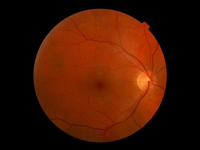
Picture of the human retina; the bright spot on the right is the blind spot where the optic nerve leaves the eye.
Steven Novella recently wrote about so-called “chiropractic neurology” and its most outspoken proponent, Ted Carrick. In 2005 I published an article in The Scientific Review of Alternative Medicine (Vol 9, No 1, p. 11-15) entitled “Blind-Spot Mapping, Cortical Function, and Chiropractic Manipulation.” It was an analysis of a study Carrick had published.
Carrick read a shorter, popularized version of my critique in Skeptical Inquirer and responded with a diatribe that was inaccurate, distorted what I had said, and accused me of fraud, deception, and mis-representation. He failed to offer a credible rebuttal of my specific criticisms; and, in my opinion, showed that he failed to understand some of my points. He referred to me as “Ms. Hall” and suggested that I was psychotic. He characterized my e-mail correspondence with him as “bizarre, rude, and offensive.” It was none of those, and I have copies of the e-mails to prove it. Carrick says he “forwarded it to the legal council for the American Chiropractic Association for review.” Now that strikes me as bizarre.
I am re-publishing the entire text of my article here as an instructive example of what passes for science in the chiropractic neurology community. Readers can judge for themselves whether my critique amounts to fraud and whether I am showing signs of psychosis, whether Carrick is a good scientist and whether his reply to my critique was appropriate.
Title: Blind-Spot Mapping, Cortical Function, and Chiropractic Manipulation
ABSTRACT:
Background: A technique of blind-spot mapping is being used by chiropractors to determine hemispheric dominance and to map cortical function; the findings are being used clinically to guide unilateral cervical spine manipulations.
Objective: The objective of this paper is to evaluate the evidence for the assertions that (1) large numbers of normal people have an enlarged blind spot on one side; (2) mapping the blind spot is equivalent to mapping brain function, and (3) spinal manipulation alters the size of the blind spot by altering brain function.
Design: A review of the literature found only 1 pertinent study; this study was analyzed for flaws and for quality of evidence.
Results: Serious methodological and logical flawswere found that invalidate the conclusions of the study.
Conclusion: The evidence for a high incidence of enlarged blind spots is unconvincing. There is no evidence that blind-spot mapping reflects cortical function or that manipulation can affect the size of the blind spot by altering brain function.
BACKGROUND
The blind spot is the area of the retina where the optic nerve enters the eye, where there are no photoreceptors. In monocular vision, it represents a small area of the visual field, about halfway between the midpoint and the lateral edge, where no visual input is received. In binocular vision, that area is visible to the opposite eye. The brain fills in the missing information to create the illusion of an uninterrupted visual field. Ophthalmologists routinely map the blind spots when testing visual fields for clinical diagnosis and follow-up. Until 1997, no one had observed any significant difference in blind-spot size between the left and right eyes in the absence of retinal disease. In that year, a study by Frederick Carrick, DC, PhD, titled “Changes in Brain Function after Manipulation of the Cervical Spine,” was published in the Journal of Manipulative and Physiological Therapeutics. (1) Studying 500 volunteers, he reported that all had a significant enlargement of 1 blind spot and that it returned to normal size following selective unilateral manipulation of the cervical spine.
The blind-spot-mapping technique described by Dr. Carrick is now being widely used by chiropractors and others. The technique is being taught at postgraduate chiropractic neurology seminars. Proponents claim to be mapping brain function, and to be able to affect brain function by manipulating the spine. A sample newspaper ad reads:
DO YOU HAVE A GOOD BRAIN OR A BAD BRAIN? ONE SIMPLE TEST MAY TELL YOu. Balance Problem? Learning Disability? Pain? Eye Problems? You name it. It could all be in your head. Researchers concluded: “Accurate reproducible maps of cortical (i.e. brain) responses can be used to measure the neurological consequences of spinal joint manipulation. Cervical manipulation activates specific neurological pathways. Manipulation of the cervical spine may be associated with an increase or a decrease in brain function depending upon the side of the manipulation and the cortical hemisphericity (i.e., side of decreased brain function) of a patient. Call Today for a FREE Brain Function Exam. (2)
Chiropractic originally claimed that all disease resulted from the effect of bone displacements (subluxations) on the nervous system. (3) Some chiropractors now limit their practice to the musculoskeletal system; others still claim to be able to treat other systems of the body. Ear infections, asthma, and constipation are among the conditions treated by chiropractors. (4) Neuroanatomy, as it is presently understood, tells us that spinal manipulation should not affect the brain or other structures within the skull; chiropractors believe it does, and they have been seeking scientific proof of that belief for over a century. The 1997 study by Dr. Carrick is considered by some to constitute that proof. The issue is an important one: if manipulation is proven to have an effect on brain function, there will be reason to accept chiropractic as a part of mainstream medicine and to teach it in medical schools.
OBJECTIVE
The evidence for blind-spot mapping was evaluated to determine if it supported claims that
- large numbers of normal people have an enlarged blind spot on one side;
- mapping the blind spot is equivalent to mapping brain function;
- spinal manipulation affects the size of the blind spot by altering brain function.
DESIGN
The medical literature and the Internet were searched and the author of the original 1997 study was contacted in an attempt to locate all published literature on the subject. Ten letters to the editor followed the original study in the Journal of Manipulative and Physiological Therapeutics, each answered by the author; no other studies were found. The author of the 1997 article knew of no other published studies since that date, although he has other studies in progress.(5)
Information on the Internet was sparse. One Web site (6) was found that mentioned the test and the results of a clinical series: “We have tested thousands of people in the past five years and have found less than 1% have normal sized blind spot maps.” No further details or documentation were provided. This Web site advertised a device for chiropractic manipulation (an activator) and was riddled with errors (such as confusing the blind spot in the eye with a driver’s blind spot). On a second Web site, (7) a chiropractor criticized Dr. Carrick for dogmatism and failing to respond to criticism about the 1997 study and criticized a third, now-defunct Web site on which a chiropractor had made statements about the blind spot not supported by anything in Dr. Carrick’s study or in the rest of the chiropractic literature. Anecdotal evidence was found indicating that a number of clinicians have also found enlarged blind spots with the test, and that the information has been clinically useful.
In the absence of any other information suitable for analysis, this analysis is based solely on the one published article.
RESULTS
Summary of the Study
The objective was “to ascertain whether manipulation of the cervical spine is associated with changes in brain function.” The subjects were 500 volunteers enrolled in postdoctoral neurology programs. Their blind spots were mapped with a simple paper-and-pencil test by 2 “blinded” examiners, and the 2 maps were judged for reproducibility by 2 “blinded” examiners who superimposed the 2 maps over a bright light. Four hundred seventy-three pairs of maps were judged reproducible; all had an enlarged blind spot on one side, with an average circumference of approximately 1.5 times the circumference of the smaller spot. In 330 subjects, the right blind spot was larger; in 143 subjects, the left one was larger. In 20 subjects with enlarged blind spots on the right, the second cervical motion segment was reduced on the right in 10 and on the left in 10. Retesting showed that after manipulation on the right side, the right blind spot was significantly smaller and the left blind spot was unchanged; after manipulation on the left side, the right blind spot was unchanged and the left blind spot was significantly larger. The remaining 453 patients were all manipulated on the same side as the enlarged blind spot; after manipulation, the enlarged blind spot was significantly smaller, while the blind spot in the other eye did not change. The study concluded:
Accurate reproducible maps of cortical (i.e. brain) responses can be used to measure the neurological consequences of spinal joint manipulation. Cervical manipulation activates specific neurological pathways. Manipulation of the cervical spine may be associated with an increase or a decrease in brain function depending upon the side of the manipulation and the cortical hemisphericity (i.e., side of decreased brain function) of a patient.
The study refers to the blind spot maps as “physiological cortical maps” and “an integer of brain activity.”
Description of the Blind-spot Test
Ophthalmologists have a variety of standardized methods for testing visual fields. The test in this study is a simple paper-and-pencil variant of the tangent-screen method:
- A piece of paper with a dot in the center is placed at eye level 28 cm from the subject’s forehead.
- The subject covers one eye and fixates on the dot with the other eye.
- The examiner moves a small target laterally from the dot to the edge of the paper; pencil marks are made where the target seems to disappear and where it seems to reappear.
- The target is placed in the area where it was invisible and moved up, down, and at 45° angles until a total of 8 dots have been mapped to delineate the borders of the blind spot.
- The 8 pencil marks are connected with straight lines and the circumference of the blind spot is measured.
Possible Methodological Flaws
- The test as described is imprecise and would be expected to vary with many factors such as degree of cooperation, speed of target movement, slight tilting of the head, reliability of fixation, judgment as to where to draw the dots, etc.
- The measurements of this new blind-spot mapping method were reproducible within the study but were not shown to be valid; there was no comparison with measurements by other, older methods.
- Reproducibility was determined by superimposing 2 drawings over a bright light; this was a subjective judgment. The maximum difference allowed between 2 “reproducible” maps was not quantified.
- The investigators may have been biased, as suggested by the objective of the study. (They were trying to study the effect of manipulation on brain function, not simply to establish a blind-spot-mapping test.)
- There were no controls who were not manipulated or who received sham manipulation or manipulation of levels below the second cervical vertebra.
- The subjects were postgraduate chiropractic neurology students. They were presumably biased in favor of chiropractic and may have been biased towards finding expected results. They knew which side of their spine had been manipulated and therefore understood which eye would be expected to show a change.
- Although the examiners were taught to monitor the subject’s eye movements, it seems unlikely that they could watch the subject’s eyes and draw dots on the paper at the same time. The subject may have been able to glance surreptitiously at how the map was developing. The subject may have been able to compare the left map to the right.
- All 500 subjects had enlarged blind spots, but the study does not mention whether anyone had equal blind spots on initial testing and was not included in the study.
References Cited in the Study
The 47 references listed are mostly irrelevant or misinterpreted and do not support the findings and conclusions of the study. Reference 1 describes how the brain chooses what illusion to put in the blind spot when adjacent signals are ambiguous, i.e., where 2 color zones abut. Reference 2 is cited to support the statement, “Cortical receptive field size varies in different individuals.” It actually says there is a rapid variation in cortical receptive-field size in response to stabilized images in the same individual. Reference 3 says: “Ocular dominance stripes … are absent from the cortical representations of the blind spot and the monocular crescent” in the Cebus monkey. Reference 4 concerns possible mediation of contour interaction phenomenon by long-range connections within the striate cortex. From this, Dr. Carrick hypothesizes that “the frequency of firing of these cortical connections affects visual perception and enlargement of the blind spot might be attributable to any mechanism that decreases the firing rate of horizontal cortical connections, hence decreasing the probability of neuronal summation.” (1, p. 537) References 5-9 all describe retinal diseases (central-retinal vein occlusion, multiple evanescent white-dot syndrome, etc.) that may cause an apparent enlargement of the blind spot as well as other areas of loss of vision. References 10-12 refer to conditions that Dr. Carrick interprets as unexplained blind-spot enlargement but actually are known to be due to retinal disease, such as acute zonal occult outer retinopathy (AZOOR). Reference 13 shows that microwave radiation can alter the size of the blind spot (by damaging tissue). Reference 14 shows that treatment of glaucoma can reduce an enlarged blind spot caused by local pressure due to glaucoma. Reference 15 shows that glaucoma can damage the retina near the blind spot. Reference 16 states that multiple events affecting vision are often related. (It reports a case of chorioretinopathy complicated by occipital lobe infarction and sinusitis in a drug abuser.)
After citing these 16 references, Dr. Carrick concludes, “It is clear that the size and shape of the blind spot is a product of neuronal activity and that this activity can be affected by a variety of processes. It was therefore chosen as a sensitive integer of brain activity that could be used to measure changes in cortical activity if other integers were manipulated.” (1, p. 539) This conclusion is not justified.
References 17-23 refer to various methods of recording blind spots (such as computerized static perimetry and multifixation campimetry), pointing out that they are more expensive than the pencil-and-paper method.
The abstract of one of these references actually states: “Statistically there was no evident difference of the size and depth of the defects of the blind spot either between the right and left visual field or among the individual age groups” (8) — in direct antithesis to the findings of Dr. Carrick’s study.
References 24-43 consist of various studies relating to the thalamus, the optic nerve and its connections, the plasticity of the visual cortex, etc. These studies say nothing about any cortical effect on the size of the blind spot or about any cortical effect of neck manipulation. Dr. Carrick uses them as departure points for imaginative speculation about how nerve signals from the neck might possibly interact with optic-nerve signals through some mechanism which has never been recognized. For example, reference 27 is titled “In Vivo Demonstration of Altered Benzodiazepine Receptor Density in Patients with Generalized Epilepsy.” He cites this to support this statement: “The amplitude of somatosensory receptor potentials will influence the frequency of firing of cerebello-thalamocortical loops that have been shown to maintain a central integrative state of cortex.” He then speculates: “Changes in the amplitude of muscle stretch receptors and joint mechanoreceptors that dictate the frequency of firing of primary afferents that contribute to cerebellothalmocortical loops should also affect the integrative cortical state, which might affect the size of the blind spot when visual afferents are maintained in a steady state.” (1, p. 540) (Emphasis added.)
References 44-47 are Dr. Carrick’s own publications describing his manipulative techniques.
Logical Flaws
- Nothing in the paper or the references it cites justifies calling a blind-spot map “a physiological cortical map” or “an integer of brain activity.” The blind spot is a fixed anatomical feature. The brain functions to fill in the blind spot but has no effect on its size.
- When a new test contradicts the findings of all previous tests, its accuracy must be suspect. Either the blind spots are the same size or they are not. Either this study is wrong or all previous observers are wrong. The test cannot be used as a measurement tool in further experiments until this dilemma is resolved.
- Carrick’s argument boils down to this: spinal manipulation must affect cortical activity because it changes the size of the blind spot, and the blind spot must represent a map of cortical activity because its size changes with spinal manipulation. Demonstrating that the size of the blind spot changes following manipulation does not prove that manipulation changes the brain or that the brain changes the blind spot, even if no other explanation is apparent. The fact that A is followed by C does not prove that A causes B and B causes C. That amounts to what has been called a logical “sillygism.”
Other Observations
In Figure 8 of the paper, a heading reads: “Blind Spots Are the Consequence of Global Brain Activity and Representative of the Anatomical Size of the Optic Disc.” This is a false statement not justified by anything in the paper.
The language of the paper is often confused and unclear. It is difficult to understand what the author means by statements such as “A constant type of environmental stimulation (vision) was used in this research to stimulate the brain through specific known neuronal pathways. The effects of this environmental stimulation were used to affect human perceptual activity and demonstrate the effectiveness of cortical mapping through comparison.” (1, p. 531)
Neuroanatomists have mapped the nervous system in great detail, and pathways that might allow spinal manipulation to affect the brain are not known to exist.
The study describes detailed statistical analysis of manipulation experiments based on a blind-spot test that has not been shown to be valid. Statistical significance is meaningless if it is based on invalid data.
CONCLUSION
Evidence supporting the use of the blind-spot-mapping test is limited to one fatally flawed study. The evidence of this study that one blind spot is larger than the other is unconvincing and contradicts all previous findings. The assertion that the size of the blind spot can change is opposed to all present anatomical and physiological knowledge. There is no evidence that blind-spot mapping reflects cortical function or that manipulation can affect the size of the blind spot by stimulating cortical function. The study fails to meet its stated objective of ascertaining whether manipulation of the cervical spine is associated with changes in brain function. The conclusions it reaches are unjustified and illogical. The paper-and-pencil blind-spot-mapping test has not been validated, and there is no reason to think that it has any clinical usefulness.
REFERENCES
- Carrick F. 1997. Changes in brain function after manipulation of the cervical spine. J Manipulative Physiol Ther. 20(8): 529-545.
- Tacoma (Washington) News Tribune, December 31,2002.
- Palmer D. The Chiropractor: Whitefish, MT: Kessinger Publishing; 1997.
- Sportelli L. 2000. Introduction to Chiropractic. 10th ed. Norwalk, IA: Practicemakers Products.
- Author? 2002. Personal communication. December 26.
- Biokinetics Health Systems. Frequently Asked Questions. Available at: http://www.biokineticshealth.com/faqs. html. Accessed December 31,2002.
- Seaman D. Philosophy and science versus dogmatism in the practice of chiropractic. The Chiropractic Resource Organization. Available at: http://www.chiro.org/LINKS/ ABSTRACTS/Science _ vs_ dogmatism.shtml. Accessed December 31, 2002.
- Lest’ak J. 1993. [Marriotte’s spot]. Cesk Oftalmol 49(6):394-398.